


Clinical AI built around how radiologists work
A medical image is acquired to answer a clinical question. A chest CT may be requested to investigate suspected pulmonary embolism. A brain MR may be performed to understand neurological symptoms. A mammogram is intended to look for signs of breast cancer. The question matters because it determines how the examination is performed, interpreted, and reported.
But the information contained in an image is not limited to the reason the examination was ordered. The same study may reveal other findings about a patient’s health, including changes that are subtle, difficult to quantify, or unrelated to the original referral. Radiologists have always considered imaging findings beyond the immediate clinical need. What is changing is the range of information that clinical AI can help make visible and measurable.
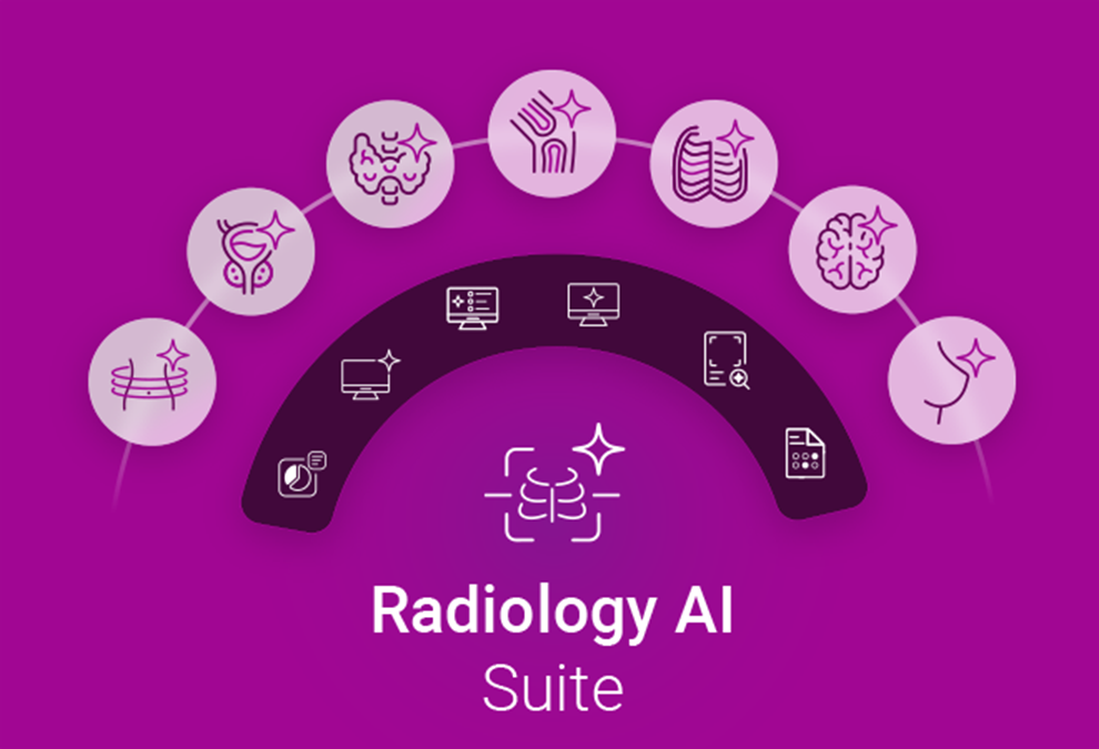
DeepHealth’s Radiology AI Suite brings together an extensive native clinical AI portfolio across imaging modalities. Its capabilities connect to the tools radiologists use to interpret images and report findings. The opportunity is to turn more of imaging’s intelligence into informed care. That raises an important question: how can healthcare make use of more information without adding unnecessary complexity to care?
What else can imaging reveal?
Consider a chest CT. Depending on the protocol and image quality, it may contain information relevant to pulmonary nodules, emphysema, and coronary artery calcification. These findings concern different clinical questions and may have different implications for follow-up.
The opportunity to address multiple clinical questions also extends to everyday chest X-ray interpretation. DeepHealth’s Chest XRay[1] solution, built on a proprietary foundation model, helps radiologists detect and localize suspected abnormalities including nodules, consolidation, and pleural abnormalities. Within one examination, the solution has the potential to draw attention to abnormalities relevant to different clinical questions. This includes those that may require urgent attention.
Assessing future risk and monitoring disease
Image-based risk prediction is another important dimension. AI-based analysis has the capability to draw on information within a mammogram to estimate a woman’s future risk of breast cancer, even when no suspicious lesion is visible on the examination. Mammography can also reveal breast arterial calcification, a marker associated with cardiovascular risk.[2] Similarly, coronary artery calcification detected incidentally on routine chest CT is an established marker of coronary atherosclerosis and is associated with an increased risk of future cardiovascular events.[3]
Imaging also helps clinicians understand how disease changes over time. In brain MR, quantitative and longitudinal analysis can reveal changes associated with neurodegenerative conditions. Together, these applications extend imaging’s contribution beyond detecting disease in the present to assessing future risk and monitoring progression, alongside clinical history and other relevant information.
The potential is meaningful, particularly as healthcare looks for opportunities to identify risk earlier and intervene when more options may be available. But it is also important to distinguish a measurable imaging feature from a validated prediction of future disease. The clinical relevance of a risk assessment depends on the evidence supporting it, the population in which it is used, and whether it can appropriately inform a decision.
Making more of the information clinically useful
More findings do not automatically mean better care. A radiologist needs to understand the evidence behind an AI-generated finding, assess it in context, and decide what belongs in the interpretation. The referring clinician needs to understand whether anything should change in the patient’s care.
This is where the experience around clinical AI becomes important. If relevant findings sit in separate applications, the radiologist may still need to locate them, reconcile measurements, and carry information manually into the report. Increasing the amount of information available can also increase the work required to use it.
A more connected approach brings clinical AI into the workflow where interpretation happens. Findings, measurements, and visualization can be made available alongside the images and clinical context. These can then be carried into reporting for the radiologist to review and refine.
The aim is to help radiologists make informed judgments while minimizing nonclinical tasks. This includes the work of assembling the intelligence around a case themselves.
Connecting clinical AI to interpretation and reporting
In thyroid ultrasound, for example, DeepHealth’s AI[4] measures and characterizes nodules and generates standardized sonographer worksheets and a pre-draft report. Radiologists can compare findings with prior studies to assess changes, then review and refine information automatically populated into the report. Measurements and findings carry forward through the examination, reducing the need to transfer information manually.
This is DeepHealth’s vision of One Intelligent Experience: relevant findings, measurements, and context brought together where radiologists review images, assess their significance, and communicate what comes next.
Performance analytics help organizations understand how that experience is working in practice. For instance, how clinical AI is being used, how it performs, whether it supports the intended workflows, and where improvement is needed. As adoption expands across clinical areas, sites, and modalities, these insights can guide improvements. This helps teams realize more value from AI in everyday care.
Radiology AI Suite brings clinical intelligence together
DeepHealth’s Radiology AI Suite brings the industry’s largest portfolio of native clinical AI together with intelligent workflow and performance analytics on one cloud-enabled platform. The suite’s clinical applications span seven areas and five core imaging modalities, supporting capabilities from triage and detection to assessment, risk prediction, and monitoring.
AI findings appear in the tools radiologists use to prioritize cases, review images, and create reports. This brings the portfolio’s clinical capabilities directly into everyday radiology practice.
DeepHealth develops and owns the clinical AI applications in its native portfolio. This gives customers a direct connection to the teams improving them. Real-world clinical feedback informs development, validation, and continued innovation.
What this means for the next era of imaging
For healthcare leaders, this means being able to address an immediate clinical need while building toward broader AI adoption. An organization can introduce applications in priority areas, connect them to existing workflows, and add capabilities as its needs evolve.
Imaging has long played a central role in answering the questions that bring patients to care. Clinical AI is opening up new ways to understand what those images may reveal, including information relevant to risk and disease progression.
The opportunity is to bring more of imaging’s intelligence into the moments when it can make a difference for clinicians and patients.
[1] DeepHealth Chest XRay, manufactured by Gleamer and distributed by DeepHealth, Inc.
[2] Nandurkar et al., “Breast Arterial Calcification as a Predictor for Future Cardiovascular Events and Mortality: A Systematic Review and Meta-analysis,” Journal of Breast Imaging, 2026.
[3] Osborne-Grinter et al., “Prevalence and clinical implications of coronary artery calcium scoring on non‑gated thoracic computed tomography: a systematic review and meta‑analysis,” European Radiology, 2024.
[4] Thyroid Ultrasound Solution includes DeepHealth Viewer and DeepHealth Thyroid US. DeepHealth Viewer is manufactured as eRAD PACS by eRAD and distributed by DeepHealth. DeepHealth Thyroid US is manufactured as See-Mode Augmented Reporting Tool, Thyroid (SMART-T) by See-Mode and distributed by DeepHealth, Inc.
Explore Radiology AI Suite